

Tendon rupture: symptoms, causes and treatments
The human body is a remarkably resilient piece of machinery, but some structures remain vulnerable to repeated strain or sudden trauma. The tendon is one of them. This band of connective tissue that links muscle to bone plays a central role in every movement of daily life, and its rupture is one of the most disabling injuries there is. A tendon rupture can happen to an elite athlete just as easily as to a sedentary person who makes an awkward move, and it requires prompt treatment to avoid lasting after-effects. Understanding how it happens, recognizing its signs and knowing the available treatment options is essential for responding appropriately.
What is a tendon rupture ?
A tendon is a dense fibrous structure, made mainly of collagen, that transmits muscle force to the bones to allow movement. Unlike muscle, it has a poor blood supply and therefore heals more slowly. There are two main types of rupture: a partial tendon tear, in which only some of the fibers are torn, and a complete tendon rupture, in which the tendon breaks entirely, interrupting all transmission of muscle force. This distinction is critically important, because it directly guides the choice of treatment and the outlook for recovery. The tendons most commonly affected are the Achilles tendon, the rotator cuff tendons in the shoulder, the patellar tendon in the knee and the flexor tendons of the hand.
Causes of a tendon rupture
A tendon rupture never really happens by chance. It is almost always the result of a triggering factor acting on tissue that was already weakened, or one that suddenly subjects the tissue to a load it cannot absorb.
The most common cause is acute trauma : a direct blow, a sudden violent muscle contraction, or stretching beyond the tendon’s elastic limits. This type of rupture is common in explosive sports such as football (soccer), basketball, tennis and track and field, where quick starts and sudden changes of direction push the tendons to their limits.
Alongside trauma, there is also a so-called degenerative form of rupture, linked to the progressive wear of tendon tissue. With age, collagen fibers lose their elasticity and strength. Untreated chronic tendinopathy can therefore lead to a spontaneous tear, sometimes with no apparent exertion. This is particularly seen in the Achilles tendon in people over 40 or in those who return to physical activity after a long period of inactivity.
Certain risk factors significantly increase the likelihood of rupture: long-term corticosteroid use, which weakens tendon tissue, certain antibiotics in the fluoroquinolone family, poor sports technique, unsuitable equipment, and obesity, which multiplies the mechanical load on weight-bearing tendons.
Symptoms to recognize
Knowing how to identify the signs of a tendon rupture can make the difference between a quick recovery and a serious complication. Symptoms vary depending on the tendon involved and the extent of the injury, but several signs consistently appear.
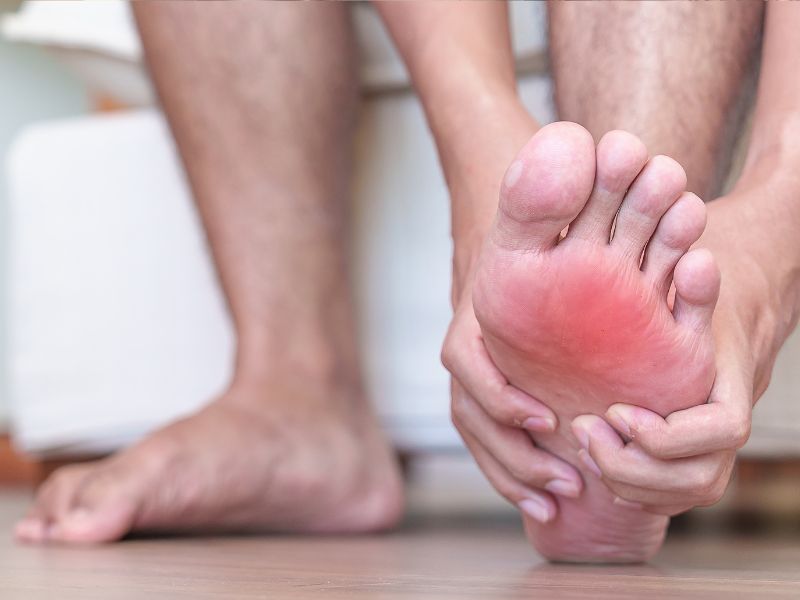
The first sign is usually sudden, intense pain, often described as a snap or a whiplash, at the moment of the injury. This pain may then ease within a few minutes, which leads some people to underestimate how serious their injury is. Localized swelling follows, along with bruising whose extent depends on the blood supply of the area involved. Loss of function is another major sign: the person can no longer perform certain specific movements, such as rising onto the toes in the case of an Achilles tendon rupture, or raising the arm in the case of a rotator cuff injury.
In complete ruptures, a dip or gap can sometimes be felt at the site of the tear, a pathognomonic sign of a break in the tendon’s continuity. Marked muscle weakness invariably accompanies this clinical picture. It is important to note that some partial ruptures can have less dramatic symptoms, which sometimes delays the diagnosis and complicates treatment.
How is the diagnosis made ?
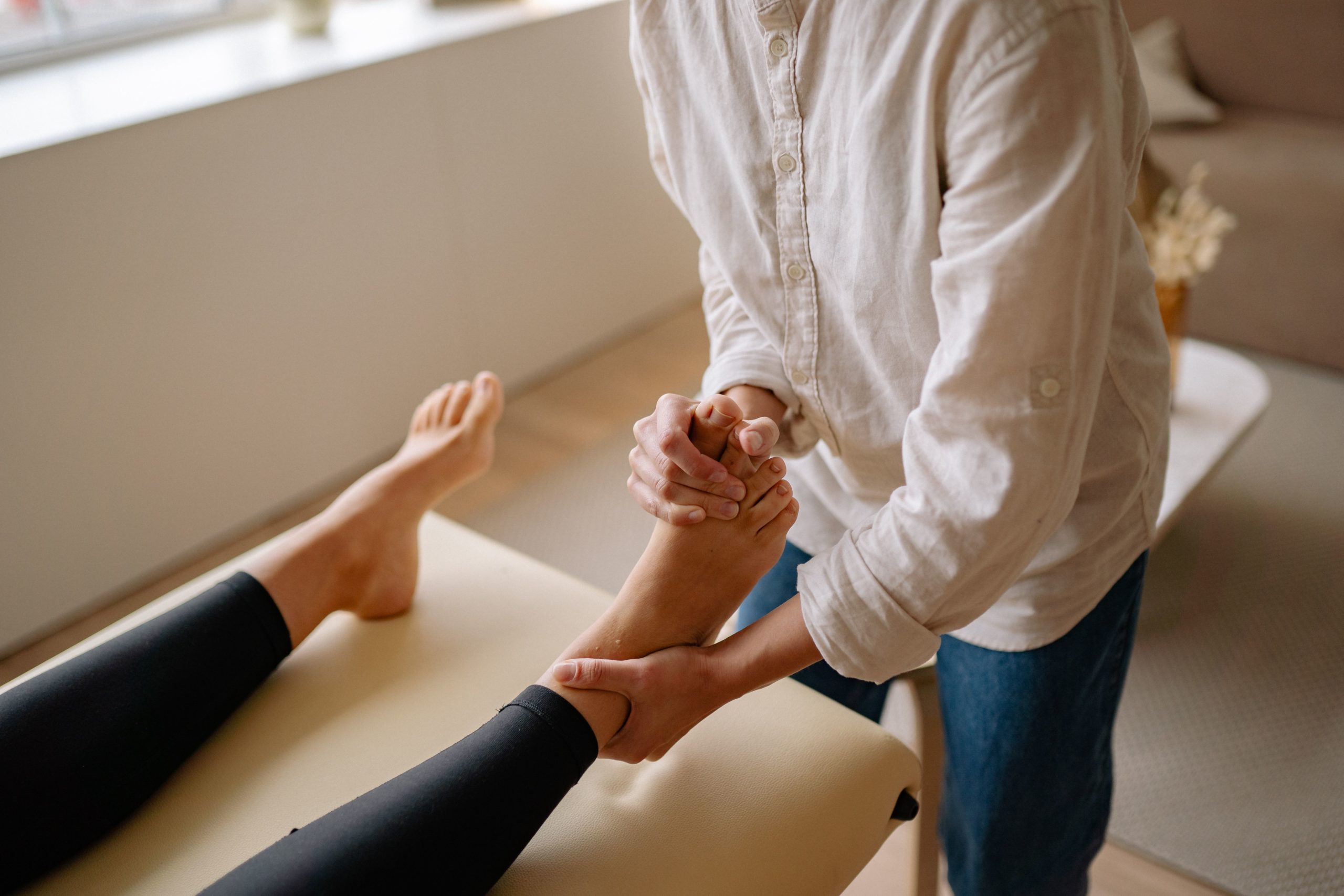
When a tendon rupture is suspected, the doctor begins with a thorough clinical examination, looking for the signs described above. Specific functional tests make it possible to assess the continuity of the tendon. For the Achilles tendon, the Thompson test, in which the calf is squeezed with the patient lying face down to see whether the foot moves, is particularly reliable.
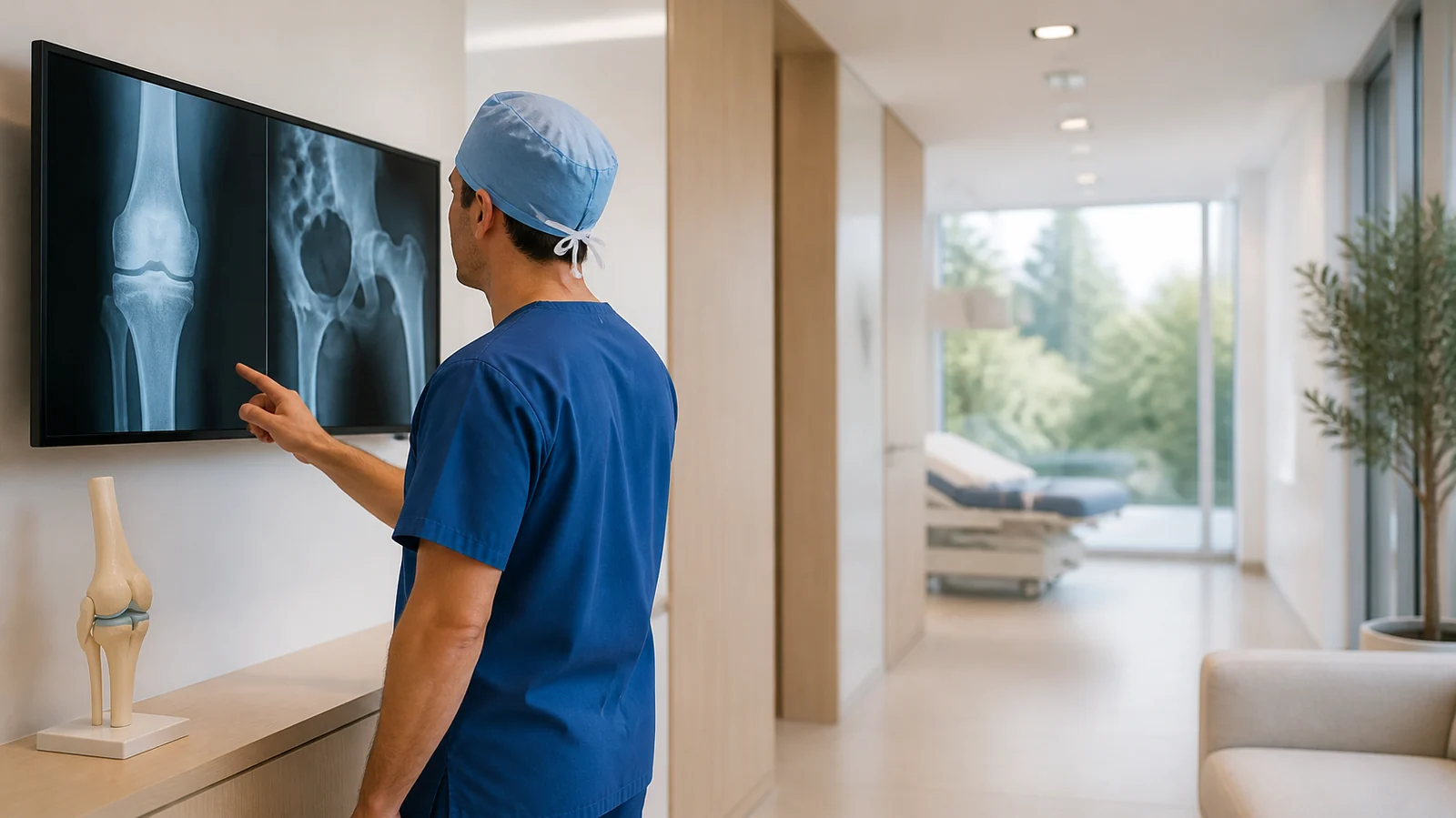
Medical imaging then confirms the diagnosis and determines its extent. A tendon ultrasound is the first-line test: quickly available, inexpensive and dynamic, it lets the doctor see the structure of the tendon in real time and assess the size of the tear. MRI (magnetic resonance imaging) is indicated in complex cases or when surgery is being considered: it gives a more precise view of the surrounding soft tissues and distinguishes a partial rupture from a complete one with great reliability. A standard X-ray, on the other hand, does not directly show tendons but can be useful for ruling out an associated fracture.
Treatment options
Management of a tendon rupture depends on several factors: the location and extent of the injury, the patient’s age, their level of physical activity and their medical history. There are two main treatment approaches, which are regularly debated in the medical literature.
Conservative (non-surgical) treatment
In some situations, particularly for partial ruptures or in patients with moderate physical activity, non-surgical treatment may be considered. It relies on immobilizing the injured area with a splint or cast, allowing the tendon fibers to regenerate gradually. This immobilization phase is followed by progressive functional rehabilitation supervised by a physical therapist. Pain relievers and anti-inflammatory drugs are prescribed to manage pain during the acute phase. This treatment requires strict compliance and a good deal of patience, because tendon healing is slow, often taking several weeks to several months.
Surgical treatment
Tendon surgery is often recommended for complete ruptures, particularly in young, active or athletic patients, because it restores the continuity of the tendon more reliably and optimizes long-term functional results. The procedure consists of suturing the two ends of the torn tendon together, sometimes using a tendon graft when the loss of tissue is too great. It is performed under regional or general anesthesia and, depending on the case, can be done as outpatient surgery. Post-operative rehabilitation is essential and is a key part of a successful outcome.
Rehabilitation: an essential step
Whichever treatment is chosen, tendon rehabilitation is the key to a full recovery. It begins with a phase of gentle mobilization aimed at limiting scar adhesions, then progresses to gradual muscle-strengthening exercises, controlled stretching and proprioceptive work to restore sensation and coordination. Total recovery time varies considerably depending on the severity of the injury and the tendon involved, but it generally ranges from three to six months, and can take up to a year for a return to high-intensity sport. Neglecting this rehabilitation phase carries a high risk of re-rupture or lasting functional limitations.
Prevention: protecting your tendons every day
While a tendon rupture can’t always be avoided, several habits can significantly reduce the risk. A proper warm-up before any physical activity, a sensible progression in training intensity, wearing shoes suited to your sport and maintaining a healthy body weight are among the most effective measures. If tendon pain persists, seeing a doctor promptly, before chronic tendinopathy progresses to a tear, is an essential precaution.
Conclusion
A tendon rupture is a serious injury that deserves treatment without delay. Recognizing its symptoms, seeing a doctor quickly and following the prescribed treatment and rehabilitation protocol to the letter are the three pillars of a successful recovery. With appropriate care, the great majority of patients regain normal function and can return to their activities, sports-related or not, under good conditions.